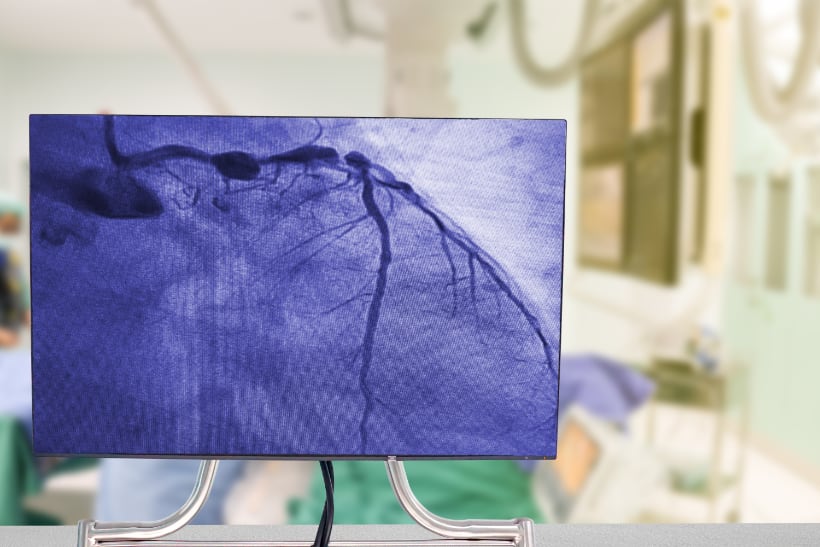
Intra-coronary imaging plays a vital role in assessing lesions and their characteristics. This allows for optimal stent selection and deployment. Thus, intuitively, this results in reduction in major adverse cardiovascular events (MACE), target lesion revascularisation (TLR) and even all-cause mortality as evidence by the IVUS-XPL, ULTIMATE and COREA-AMI data1,2,3.
The RENOVATE-COMPLEX-PCI trial was a randomised, multi-centre, open-label trial assessing differences in outcomes in patients with complex coronary disease undergoing intracoronary imaging guided percutaneous coronary intervention (PCI) versus angiographically guided PCI.
Complex coronary anatomy was defined as: true bifurcation lesions with a side branch diameter > 2.5mm; chronic total occlusion (CTO); unprotected left main stem; a stent length expected to be > 38mm; multi vessel or multi stent PCI; in stent restenosis, ostial lesions and severely calcified lesions. Patients with cardiogenic shock, intolerance to anti thrombotic therapies and anaphylactic reactions to contrast dye were excluded.
A total of 1,639 patients were randomised in a 2:1 fashion to intracoronary imaging guided PCI (n=1092) and angiographic guided PCI (n=547). The mean age of the patients were 66 years of age with 79% being male. Just over a third (38%) had diabetes and 51% of PCI was completed in the setting of an acute coronary syndrome (51%). In the intra-coronary imaging arm, Intravascular ultrasound (IVUS) was utilised in 75% of patients whilst the remaining 25% underwent imaging via optical coherence tomography (OCT). Patients were followed up for 3 years.
At a median duration of 2.1 years, the primary composite endpoint of cardiac mortality, target-vessel-related myocardial infarction or clinically driven target vessel revascularisation was significantly reduced in the intracoronary imaging cohort (7.7%) when compared to the angiography arm (12.3%) (HR 0.64, 95% CI 0.45-0.89, P=0.008). Interestingly, this difference was primarily due to a significant reduction in mortality from cardiac causes (1.7% vs 3.8%, HR 0.47, 95% CI 0.24-0.93). In a sensivity analysis which excluded peri-procedural myocardial infarction, the primary endpoint was still significantly reduced in the intra-coronary imaging arm. However, both the rates of myocardial infarction and repeat revascularisation did not statistically differ between both arms but there was a signal towards enhanced outcomes with intra-coronary imaging.
This study provides more data demonstrating the consistent benefits of intracoronary imaging guided PCI versus angiographic guided PCI over a 3-year follow-up period. The mechanisms by which cardiac mortality is significantly reduced are not clear but the numerical trend towards better outcomes in myocardial infarction and repeat revascularisation may provide the answers to this prognostic benefit. It is overwhelmingly evident that in complex coronary artery disease intracoronary imaging should be employed. Consequently, the routine use of intracoronary imaging in all-comer coronary disease is something that needs to be strongly considered before the deployment of stents.
Link to article – https://www.nejm.org/doi/full/10.1056/NEJMoa2216607
References
- Hong, S.J., Kim, B.K., Shin, D.H., Nam, C.M., Kim, J.S., Ko, Y.G., Choi, D., Kang, T.S., Kang, W.C., Her, A.Y. and Kim, Y.H., 2015. Effect of intravascular ultrasound–guided vs angiography-guided everolimus-eluting stent implantation: the IVUS-XPL randomized clinical trial. Jama, 314(20), pp.2155-2163.
- Zhang, J., Gao, X., Kan, J., Ge, Z., Han, L., Lu, S., Tian, N., Lin, S., Lu, Q., Wu, X. and Li, Q., 2018. Intravascular ultrasound versus angiography-guided drug-eluting stent implantation: the ULTIMATE trial. Journal of the American College of Cardiology, 72(24), pp.3126-3137.
- Choi, I.J., Lim, S., Choo, E.H., Hwang, B.H., Kim, C.J., Park, M.W., Lee, J.M., Park, C.S., Kim, H.Y., Yoo, K.D. and Jeon, D.S., 2021. Impact of intravascular ultrasound on long-term clinical outcomes in patients with acute myocardial infarction. Cardiovascular Interventions, 14(22), pp.2431-2443.

